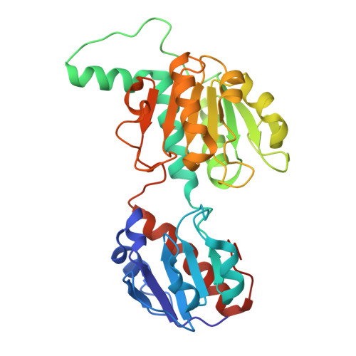
9IBE | pdb_00009ibe

D-2-hydroxyacid dehydrogenase (D2HDH) from Haloferax mediterranei in complex with potassium, 2-ketohexanoic acid, NADP+ and chloride


Experimental Data Snapshot

  • Method: X-RAY DIFFRACTION
  • Resolution: 1.26 Å
  • R-Value Free: 
    0.188 (Depositor), 0.192 (DCC) 
  • R-Value Work: 
    0.152 (Depositor), 0.160 (DCC) 

Starting Model: experimental
View more details

wwPDB Validation   3D Report Full Report


Ligand Structure Quality Assessment 


This is version 1.0 of the entry. See complete history


Literature

Potassium binding by carbonyl clusters, halophilic adaptation and catalysis of Haloferax mediterranei D-2-hydroxyacid dehydrogenase.

Domenech, J.Pramanpol, N.Bisson, C.Sedelnikova, S.E.Barrett, J.R.Dakhil, A.A.A.B.Mykhaylyk, V.Abdelhameed, A.S.Harding, S.E.Rice, D.W.Baker, P.J.Ferrer, J.

(2025) Commun Biol 8: 1170-1170

  • DOI: https://doi.org/10.1038/s42003-025-08587-7
  • Primary Citation of Related Structures:  
    5MH5, 5MH6, 5MHA, 8QZA, 8QZB, 9IBE

  • PubMed Abstract: 

    Enzymes from salt-in halophiles are stable in conditions of low water activity with applications in chiral synthesis requiring organic solvents, yet the origins of such stability remains poorly understood. Here we describe the molecular basis of the reaction mechanism and dual NADH/NADPH-specificity of D2HDH, a 2-hydroxyacid dehydrogenase from the extreme halophile Haloferax mediterranei, an organism whose proteins have to remain active in high intracellular concentrations of KCl. Halophilic adaptations of D2HDH include the expected acidic surface and a reduction in hydrophobic surface resulting from a lower lysine content. Structure determination of crystals of D2HDH grown with KCl showed that bound K + ions were coordinated predominantly by clusters of main chain protein carbonyl ligands, with no involvement of the numerous exposed surface carboxyls. Structural comparisons identified similar sites in other halophilic proteins suggesting that the generic use of carbonyl clusters to coordinate K + ions may also contribute in a carboxylate-independent way to the stabilisation of the folded state of the protein in its high salt environment.


  • Organizational Affiliation
    • Dept. Bioquımica y Biología Molecular y EQA. Universidad de Alicante, Alicante, Spain.

Macromolecules
Find similar proteins by:  (by identity cutoff)  |  3D Structure
Entity ID: 1
MoleculeChains Sequence LengthOrganismDetailsImage
D-2-hydroxyacid dehydrogenase
A, B, C, D
308Haloferax mediterraneiMutation(s): 0 
Gene Names: ddh
EC: 1.1.1
UniProt
Find proteins for Q2VEQ7 (Haloferax mediterranei (strain ATCC 33500 / DSM 1411 / JCM 8866 / NBRC 14739 / NCIMB 2177 / R-4))
Explore Q2VEQ7 
Go to UniProtKB:  Q2VEQ7
Entity Groups  
Sequence Clusters30% Identity50% Identity70% Identity90% Identity95% Identity100% Identity
UniProt GroupQ2VEQ7
Sequence Annotations
Expand
  • Reference Sequence
Small Molecules
Ligands 5 Unique
IDChains Name / Formula / InChI Key2D Diagram3D Interactions
NAP (Subject of Investigation/LOI)
Query on NAP

Download Ideal Coordinates CCD File 
DA [auth C],
E [auth A],
QA [auth D],
R [auth B]
NADP NICOTINAMIDE-ADENINE-DINUCLEOTIDE PHOSPHATE
C21 H28 N7 O17 P3
XJLXINKUBYWONI-NNYOXOHSSA-N
7N5 (Subject of Investigation/LOI)
Query on 7N5

Download Ideal Coordinates CCD File 
EA [auth C],
F [auth A],
RA [auth D],
S [auth B]
2-Ketohexanoic acid
C6 H10 O3
XNIHZNNZJHYHLC-UHFFFAOYSA-N
K (Subject of Investigation/LOI)
Query on K

Download Ideal Coordinates CCD File 
FA [auth C]
G [auth A]
GA [auth C]
H [auth A]
HA [auth C]
FA [auth C],
G [auth A],
GA [auth C],
H [auth A],
HA [auth C],
I [auth A],
IA [auth C],
J [auth A],
JA [auth C],
K [auth A],
KA [auth C],
L [auth A],
LA [auth C],
M [auth A],
SA [auth D],
T [auth B],
TA [auth D],
U [auth B],
UA [auth D],
V [auth B],
VA [auth D],
W [auth B],
X [auth B]
POTASSIUM ION
K
NPYPAHLBTDXSSS-UHFFFAOYSA-N
CL (Subject of Investigation/LOI)
Query on CL

Download Ideal Coordinates CCD File 
CA [auth B],
P [auth A],
PA [auth C],
Q [auth A],
ZA [auth D]
CHLORIDE ION
Cl
VEXZGXHMUGYJMC-UHFFFAOYSA-M
MG
Query on MG

Download Ideal Coordinates CCD File 
AA [auth B]
BA [auth B]
MA [auth C]
N [auth A]
NA [auth C]
AA [auth B],
BA [auth B],
MA [auth C],
N [auth A],
NA [auth C],
O [auth A],
OA [auth C],
WA [auth D],
XA [auth D],
Y [auth B],
YA [auth D],
Z [auth B]
MAGNESIUM ION
Mg
JLVVSXFLKOJNIY-UHFFFAOYSA-N
Experimental Data & Validation

Experimental Data

  • Method: X-RAY DIFFRACTION
  • Resolution: 1.26 Å
  • R-Value Free:  0.188 (Depositor), 0.192 (DCC) 
  • R-Value Work:  0.152 (Depositor), 0.160 (DCC) 
Space Group: P 1
Unit Cell:
Length ( Å )Angle ( ˚ )
a = 66.633α = 108.841
b = 75.34β = 108.084
c = 78.234γ = 95.526
Software Package:
Software NamePurpose
REFMACrefinement
xia2data reduction
DIALSdata scaling
PHASERphasing

Structure Validation

View Full Validation Report


Ligand Structure Quality Assessment 


Entry History & Funding Information

Deposition Data


Funding OrganizationLocationGrant Number
Biotechnology and Biological Sciences Research Council (BBSRC)United KingdomBB/1003703/1
Biotechnology and Biological Sciences Research Council (BBSRC)United KingdomBB/D524975/1
Generalitat ValencianaSpainGV05/166

Revision History  (Full details and data files)

  • Version 1.0: 2025-08-20
    Type: Initial release