Uncovering druggable hotspots on Pin1 via X-ray crystallographic fragment screening.
Xiao, Q., Tang, J., Shu, H., Wu, T., Zhang, H., Wang, W., Wang, L., Qin, W.(2025) Eur J Med Chem 299: 118048-118048
- PubMed: 40803165
- DOI: https://doi.org/10.1016/j.ejmech.2025.118048
- Primary Citation of Related Structures:
9JYO, 9JYP, 9JYR, 9JYT, 9JYV, 9JZ2, 9JZ3, 9JZ4, 9JZ6, 9JZG, 9JZS, 9JZU, 9JZV, 9KE7, 9KE9, 9KEB, 9KEC, 9KEL, 9KEQ, 9KES, 9KEW, 9KEY, 9KEZ, 9KF0, 9KFC, 9KFH, 9KG9, 9KX7, 9KX9, 9KXC, 9KXD, 9KXE, 9KXF, 9KXG, 9KXH, 9KXI, 9KXJ, 9KXL, 9KXM, 9KXN, 9KXO, 9KXP, 9KXQ, 9V92 - PubMed Abstract:
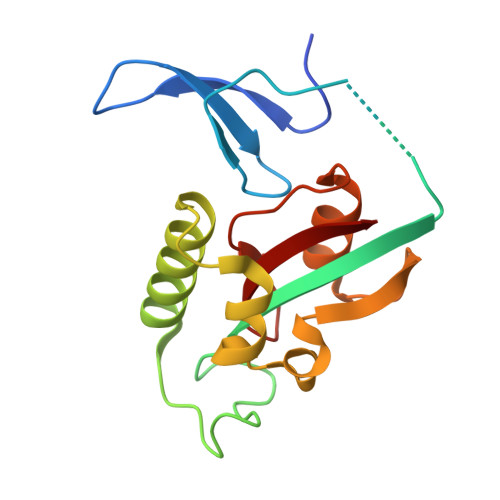
Pin1 is a phosphorylation-dependent peptidyl-prolyl isomerase that specifically recognizes and catalyzes the cis-trans isomerization of pSer/Thr-Pro motifs. It plays a pivotal role in cell cycle regulation, signal transduction, and tumorigenesis. Due to its overexpression in many cancer types, Pin1 has emerged as a promising target for the development of anticancer drugs. However, the relatively shallow and flat surface of Pin1 presents significant challenges for traditional small-molecule inhibitor design. To overcome these limitations, we employed an X-ray crystallography-based fragment screening strategy and identified approximately 50 Pin1-fragment complex structures from a curated fragment library. Systematic structural analysis revealed several druggable binding hotspots, including the well-characterized catalytic center (Site 1) and a neighboring region near the catalytic residue Cys113 (Site 2). Both sites supported stable binding with diverse fragment scaffolds. Notably, several fragments displayed cooperative binding across multiple sites, highlighting their potential as scaffolds for multifunctional inhibitor design. Additionally, a subset of fragments showed reactivity toward Cys113, and their covalent binding modes were confirmed through crystallographic and mass spectrometric analyses. Enzymatic inhibition assays further demonstrated that several fragments effectively suppressed Pin1 activity in solution, validating their potential as lead compounds. In summary, this study systematically mapped functional binding pockets on Pin1 through a structure-driven fragment screening approach, expanded its druggable landscape, and identified key structural features and fragment chemotypes to guide the development of selective, well-defined Pin1 inhibitors.
- National Facility for Protein Science in Shanghai, Shanghai Advanced Research Institute, Chinese Academy of Sciences, Shanghai, 201210, People's Republic of China.
Organizational Affiliation: