9MVK | pdb_00009mvk

Co-crystal structure of feline coronavirus UU23 main protease with nirmatrelvir


Experimental Data Snapshot

  • Method: X-RAY DIFFRACTION
  • Resolution: 1.97 Å
  • R-Value Free: 
    0.231 (Depositor), 0.244 (DCC) 
  • R-Value Work: 
    0.188 (Depositor), 0.201 (DCC) 
  • R-Value Observed: 
    0.205 (Depositor) 

Starting Model: experimental
View more details

wwPDB Validation   3D Report Full Report


Ligand Structure Quality Assessment 


This is version 1.1 of the entry. See complete history


Literature

Structural Analysis of Inhibitor Binding to the Feline Enteric Coronavirus (FECV) Main Protease.

Maryam, A.Moquin, S.A.Dovala, D.Kaur, J.Kurt Yilmaz, N.Shaqra, A.M.Schiffer, C.A.

(2025) Viruses 17

  • DOI: https://doi.org/10.3390/v17111506
  • Primary Citation of Related Structures:  
    9MVK, 9MVL, 9MW4, 9PQG

  • PubMed Abstract: 

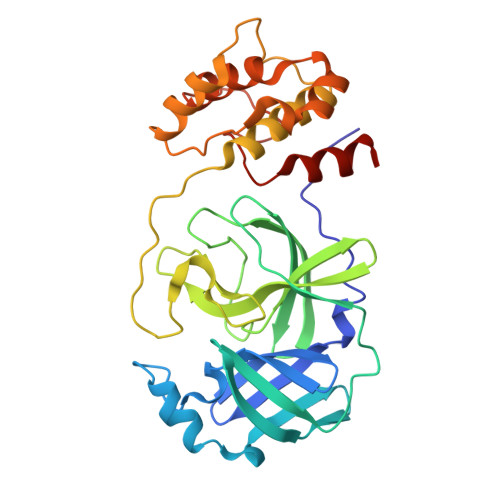
    Coronaviruses include various strains that reside in natural animal reservoirs, with zoonotic transmission posing risks to both domesticated animals and human health. Recent efforts to address coronavirus infections have focused on developing inhibitors targeting the main protease (M pro ), some of which exhibit potential broad-spectrum efficacy. This study presents crystal structures of four clinically relevant inhibitors-GC376, PF-00835231, nirmatrelvir, and ibuzatrelvir-bound to M pro from the feline coronavirus strain FECV-UU23. Structural analysis identified distinct FECV-specific features within the active site where these inhibitors bind and revealed S4 loop as a susceptible structural region essential for the enhanced binding of inhibitors in UU23 M pro . We therefore propose to incorporate sterically constrained, functionally tailored heterocyclic moieties at the P3 site of known inhibitors which can optimally engage Q187, P188, and S189 residues of the S4 loop. The findings presented enhance understanding of inhibitor specificity and reinforce the promise of these inhibitor scaffolds for developing antivirals against feline coronavirus strains, with possible applications in broad-spectrum coronavirus therapy.


  • Organizational Affiliation
    • Department of Biochemistry and Molecular Biotechnology, University of Massachusetts Chan Medical School, Worcester, MA 01605, USA.

Macromolecules
Find similar proteins by:  (by identity cutoff)  |  3D Structure
Entity ID: 1
MoleculeChains Sequence LengthOrganismDetailsImage
Peptidase C30
A, B, C, D
302Feline coronavirus UU23Mutation(s): 0 
UniProt
Find proteins for D3KDL2 (Feline coronavirus UU23)
Explore D3KDL2 
Go to UniProtKB:  D3KDL2
Entity Groups  
Sequence Clusters30% Identity50% Identity70% Identity90% Identity95% Identity100% Identity
UniProt GroupD3KDL2
Sequence Annotations
Expand
  • Reference Sequence
Experimental Data & Validation

Experimental Data

  • Method: X-RAY DIFFRACTION
  • Resolution: 1.97 Å
  • R-Value Free:  0.231 (Depositor), 0.244 (DCC) 
  • R-Value Work:  0.188 (Depositor), 0.201 (DCC) 
  • R-Value Observed: 0.205 (Depositor) 
Space Group: P 1 21 1
Unit Cell:
Length ( Å )Angle ( ˚ )
a = 63.919α = 90
b = 106.316β = 90.51
c = 78.509γ = 90
Software Package:
Software NamePurpose
PHENIXrefinement
Cootmodel building
XDSdata reduction
XDSdata scaling
PHASERphasing

Structure Validation

View Full Validation Report


Ligand Structure Quality Assessment 


Entry History & Funding Information

Deposition Data


Funding OrganizationLocationGrant Number
Other private--

Revision History  (Full details and data files)

  • Version 1.0: 2025-11-26
    Type: Initial release
  • Version 1.1: 2025-12-10
    Changes: Database references