9NY8 | pdb_00009ny8

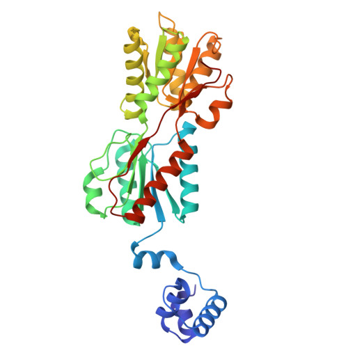
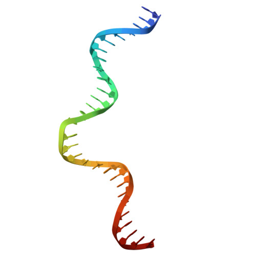
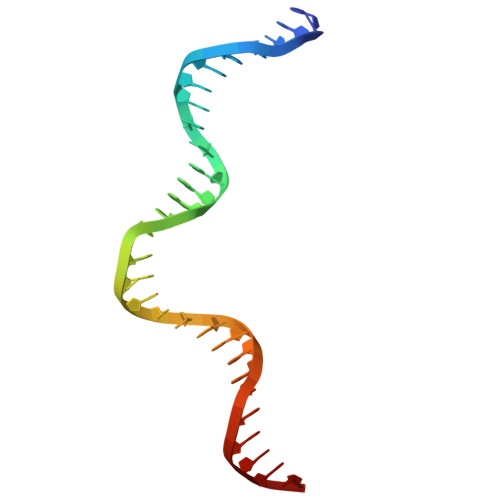
Crystal structure of the ribose operon repressor, RbsR, bound to ribose operon


Experimental Data Snapshot

  • Method: X-RAY DIFFRACTION
  • Resolution: 2.10 Å
  • R-Value Free: 
    0.223 (Depositor), 0.226 (DCC) 
  • R-Value Work: 
    0.192 (Depositor), 0.196 (DCC) 
  • R-Value Observed: 
    0.194 (Depositor) 

Starting Model: in silico
View more details

wwPDB Validation   3D Report Full Report


Ligand Structure Quality Assessment 


This is version 1.0 of the entry. See complete history


Literature

Conserved energetic changes drive function in an ancient protein fold.

Wells, M.L.Lu, C.Sultanov, D.Weber, K.C.Gong, Z.Glasgow, A.

(2025) bioRxiv 

  • DOI: https://doi.org/10.1101/2025.04.02.646877
  • Primary Citation of Related Structures:  
    9NY7, 9NY8

  • PubMed Abstract: 

    Many protein sequences occupy similar three-dimensional structures known as protein folds. In nature, protein folds are well-conserved over the course of evolution, such that there are 100,000 times as many extant protein sequences than there are folds. Despite their common shapes, similar protein folds can adopt wide-ranging functions, raising the question: are protein folds so strongly conserved for the purpose of maintaining function-driving energetic changes in protein families? Here we show that a set of key energetic relationships in a family of bacterial transcription factors (TFs) is conserved using high-resolution hydrogen exchange/mass spectrometry, bioinformatics, X-ray crystallography, and molecular dynamics simulations. We compared the TFs to their anciently diverged structural homologs, the periplasmic binding proteins (PBPs), expecting that protein families that share the same fold and bind the same sugars would have similar energetic responses. Surprisingly, our findings reveal the opposite: the "energetic blueprints" of the PBPs and the TFs are largely distinct, with the allosteric network of the TFs evolving specifically to support the functional requirements of genome regulation, versus conserved interactions with membrane transport machinery in PBPs. These results demonstrate how the same fold can be adapted for different sense/response functions via family-specific energetic requirements - even when responding to the same chemical trigger. Understanding the evolutionarily conserved energetic blueprint for a protein family provides a roadmap for designing functional proteins de novo , and will help us treat aberrant protein behavior in conserved domains in disease-related drug targets, where engineering selectivity is challenging.


Macromolecules

Find similar proteins by:  (by identity cutoff)  |  3D Structure
Entity ID: 1
MoleculeChains Sequence LengthOrganismDetailsImage
Ribose operon repressor
A, B
330Escherichia coliMutation(s): 0 
Gene Names: rbsRb3753JW3732
UniProt
Find proteins for P0ACQ0 (Escherichia coli (strain K12))
Explore P0ACQ0 
Go to UniProtKB:  P0ACQ0
Entity Groups  
Sequence Clusters30% Identity50% Identity70% Identity90% Identity95% Identity100% Identity
UniProt GroupP0ACQ0
Sequence Annotations
Expand
  • Reference Sequence
Find similar nucleic acids by:  (by identity cutoff)  |  3D Structure
Entity ID: 2
MoleculeChains LengthOrganismImage
ribose operon30Escherichia coli
Sequence Annotations
Expand
  • Reference Sequence
Find similar nucleic acids by:  (by identity cutoff)  |  3D Structure
Entity ID: 3
MoleculeChains LengthOrganismImage
ribose operon30Escherichia coli
Sequence Annotations
Expand
  • Reference Sequence
Experimental Data & Validation

Experimental Data

  • Method: X-RAY DIFFRACTION
  • Resolution: 2.10 Å
  • R-Value Free:  0.223 (Depositor), 0.226 (DCC) 
  • R-Value Work:  0.192 (Depositor), 0.196 (DCC) 
  • R-Value Observed: 0.194 (Depositor) 
Space Group: P 31 2 1
Unit Cell:
Length ( Å )Angle ( ˚ )
a = 125.53α = 90
b = 125.53β = 90
c = 122.31γ = 120
Software Package:
Software NamePurpose
REFMACrefinement
xia2data reduction
STARANISOdata scaling
Aimlessdata scaling
PHASERphasing

Structure Validation

View Full Validation Report


Ligand Structure Quality Assessment 


Entry History & Funding Information

Deposition Data


Funding OrganizationLocationGrant Number
National Institutes of Health/National Institute of General Medical Sciences (NIH/NIGMS)United StatesR00GM135529

Revision History  (Full details and data files)

  • Version 1.0: 2025-07-16
    Type: Initial release