9FMR | pdb_00009fmr

Structure of DDB1/Cdk12/Cyclin K with molecular glue SR-4835


Experimental Data Snapshot

  • Method: X-RAY DIFFRACTION
  • Resolution: 3.90 Å
  • R-Value Free: 
    0.250 (Depositor), 0.247 (DCC) 
  • R-Value Work: 
    0.208 (Depositor), 0.208 (DCC) 
  • R-Value Observed: 
    0.209 (Depositor) 

Starting Models: experimental
View more details

wwPDB Validation   3D Report Full Report


Ligand Structure Quality Assessment 


This is version 1.1 of the entry. See complete history


Literature

Discovery and design of molecular glue enhancers of CDK12-DDB1 interactions for targeted degradation of cyclin K.

Ghosh, P.Schmitz, M.Pandurangan, T.Zeleke, S.T.Chan, S.C.Mosior, J.Sun, L.Palve, V.Grassie, D.Anand, K.Frydman, S.Roush, W.R.Schonbrunn, E.Geyer, M.Duckett, D.Monastyrskyi, A.

(2024) RSC Chem Biol 6: 36-55

  • DOI: https://doi.org/10.1039/d4cb00190g
  • Primary Citation of Related Structures:  
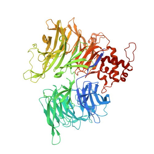
    9FMR

  • PubMed Abstract: 

    The CDK12 inhibitor SR-4835 promotes the proteasomal degradation of cyclin K, contingent on the presence of CDK12 and the CUL4-RBX1-DDB1 E3 ligase complex. The inhibitor displays molecular glue activity, which correlates with its enhanced ability to inhibit cell growth. This effect is achieved by facilitating the formation of a ternary complex that requires the small molecule SR-4835, CDK12, and the adaptor protein DDB1, leading to the subsequent ubiquitination and degradation of cyclin K. We have successfully solved the structure of the ternary complex, enabling the de novo design of molecular glues that transform four different CDK12 scaffold inhibitors, including the clinical pan-CDK inhibitor dinaciclib, into cyclin K degraders. These results not only deepen our understanding of CDK12's role in cell regulation but also underscore significant progress in designing molecular glues for targeted protein degradation in cancers associated with dysregulated cyclin K activity.


  • Organizational Affiliation

    Department of Drug Discovery, Moffitt Cancer Center Tampa Florida 33612 USA andrii.monastyrskyi@moffitt.org.


Macromolecules
Find similar proteins by:  (by identity cutoff)  |  3D Structure
Entity ID: 1
MoleculeChains Sequence LengthOrganismDetailsImage
DNA damage-binding protein 1
A, D, G
864Homo sapiensMutation(s): 0 
Gene Names: DDB1XAP1
UniProt & NIH Common Fund Data Resources
Find proteins for Q16531 (Homo sapiens)
Explore Q16531 
Go to UniProtKB:  Q16531
PHAROS:  Q16531
GTEx:  ENSG00000167986 
Entity Groups  
Sequence Clusters30% Identity50% Identity70% Identity90% Identity95% Identity100% Identity
UniProt GroupQ16531
Sequence Annotations
Expand
  • Reference Sequence
Find similar proteins by:  (by identity cutoff)  |  3D Structure
Entity ID: 2
MoleculeChains Sequence LengthOrganismDetailsImage
Cyclin-dependent kinase 12
B, E, H
351Homo sapiensMutation(s): 0 
Gene Names: CDK12CRK7CRKRSKIAA0904
EC: 2.7.11.22 (PDB Primary Data), 2.7.11.23 (PDB Primary Data)
UniProt & NIH Common Fund Data Resources
Find proteins for Q9NYV4 (Homo sapiens)
Explore Q9NYV4 
Go to UniProtKB:  Q9NYV4
PHAROS:  Q9NYV4
GTEx:  ENSG00000167258 
Entity Groups  
Sequence Clusters30% Identity50% Identity70% Identity90% Identity95% Identity100% Identity
UniProt GroupQ9NYV4
Sequence Annotations
Expand
  • Reference Sequence
Find similar proteins by:  (by identity cutoff)  |  3D Structure
Entity ID: 3
MoleculeChains Sequence LengthOrganismDetailsImage
Cyclin-K
C, F, I
268Homo sapiensMutation(s): 0 
Gene Names: CCNKCPR4
UniProt & NIH Common Fund Data Resources
Find proteins for O75909 (Homo sapiens)
Explore O75909 
Go to UniProtKB:  O75909
PHAROS:  O75909
GTEx:  ENSG00000090061 
Entity Groups  
Sequence Clusters30% Identity50% Identity70% Identity90% Identity95% Identity100% Identity
UniProt GroupO75909
Sequence Annotations
Expand
  • Reference Sequence
Small Molecules
Modified Residues  1 Unique
IDChains TypeFormula2D DiagramParent
TPO
Query on TPO
B, E, H
L-PEPTIDE LINKINGC4 H10 N O6 PTHR
Experimental Data & Validation

Experimental Data

  • Method: X-RAY DIFFRACTION
  • Resolution: 3.90 Å
  • R-Value Free:  0.250 (Depositor), 0.247 (DCC) 
  • R-Value Work:  0.208 (Depositor), 0.208 (DCC) 
  • R-Value Observed: 0.209 (Depositor) 
Space Group: P 31 2 1
Unit Cell:
Length ( Å )Angle ( ˚ )
a = 249.521α = 90
b = 249.521β = 90
c = 218.535γ = 120
Software Package:
Software NamePurpose
PHENIXrefinement
PHENIXrefinement
XDSdata reduction
XDSdata scaling
PHASERphasing

Structure Validation

View Full Validation Report


Ligand Structure Quality Assessment 


Entry History & Funding Information

Deposition Data


Funding OrganizationLocationGrant Number
German Research Foundation (DFG)GermanyEXC2151-390873048

Revision History  (Full details and data files)

  • Version 1.0: 2024-10-16
    Type: Initial release
  • Version 1.1: 2025-04-30
    Changes: Database references