2O78 | pdb_00002o78

Tyrosine ammonia-lyase from Rhodobacter sphaeroides (His89Phe variant) complexed with cinnamic acid


Experimental Data Snapshot

  • Method: X-RAY DIFFRACTION
  • Resolution: 1.90 Å
  • R-Value Free: 
    0.196 (Depositor), 0.190 (DCC) 
  • R-Value Work: 
    0.172 (Depositor), 0.170 (DCC) 

Starting Model: experimental
View more details

wwPDB Validation   3D Report Full Report


This is version 2.1 of the entry. See complete history


Literature

Structural determinants and modulation of substrate specificity in phenylalanine-tyrosine ammonia-lyases.

Louie, G.V.Bowman, M.E.Moffitt, M.C.Baiga, T.J.Moore, B.S.Noel, J.P.

(2006) Chem Biol 13: 1327-1338

  • DOI: https://doi.org/10.1016/j.chembiol.2006.11.011
  • Primary Citation of Related Structures:  
    2O6Y, 2O78, 2O7B, 2O7D, 2O7E, 2O7F

  • PubMed Abstract: 

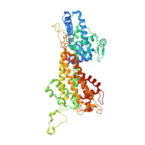
    Aromatic amino acid ammonia-lyases catalyze the deamination of L-His, L-Phe, and L-Tyr, yielding ammonia plus aryl acids bearing an alpha,beta-unsaturated propenoic acid. We report crystallographic analyses of unliganded Rhodobacter sphaeroides tyrosine ammonia-lyase (RsTAL) and RsTAL bound to p-coumarate and caffeate. His 89 of RsTAL forms a hydrogen bond with the p-hydroxyl moieties of coumarate and caffeate. His 89 is conserved in TALs but replaced in phenylalanine ammonia-lyases (PALs) and histidine ammonia-lyases (HALs). Substitution of His 89 by Phe, a characteristic residue of PALs, yields a mutant with a switch in kinetic preference from L-Tyr to L-Phe. Structures of the H89F mutant in complex with the PAL product, cinnamate, or the PAL-specific inhibitor, 2-aminoindan-2-phosphonate (AIP), support the role of position 89 as a specificity determinant in the family of aromatic amino acid ammonia-lyases and aminomutases responsible for beta-amino acid biosynthesis.


  • Organizational Affiliation
    • Howard Hughes Medical Institute, Jack H. Skirball Center for Chemical Biology and Proteomics, The Salk Institute for Biological Studies, La Jolla, California 92037, USA.

Macromolecules
Find similar proteins by:  (by identity cutoff)  |  3D Structure
Entity ID: 1
MoleculeChains Sequence LengthOrganismDetailsImage
Putative histidine ammonia-lyase
A, B, C, D, E
A, B, C, D, E, F, G, H
521Cereibacter sphaeroidesMutation(s): 1 
Gene Names: hutH
EC: 4.3.1 (PDB Primary Data), 4.3.1.23 (UniProt)
UniProt
Find proteins for Q3IWB0 (Cereibacter sphaeroides (strain ATCC 17023 / DSM 158 / JCM 6121 / CCUG 31486 / LMG 2827 / NBRC 12203 / NCIMB 8253 / ATH 2.4.1.))
Explore Q3IWB0 
Go to UniProtKB:  Q3IWB0
Entity Groups  
Sequence Clusters30% Identity50% Identity70% Identity90% Identity95% Identity100% Identity
UniProt GroupQ3IWB0
Sequence Annotations
Expand
  • Reference Sequence
Small Molecules
Modified Residues  1 Unique
IDChains TypeFormula2D DiagramParent
MDO
Query on MDO
A, B, C, D, E
A, B, C, D, E, F, G, H
L-PEPTIDE LINKINGC8 H11 N3 O3ALA, SER, GLY
Experimental Data & Validation

Experimental Data

  • Method: X-RAY DIFFRACTION
  • Resolution: 1.90 Å
  • R-Value Free:  0.196 (Depositor), 0.190 (DCC) 
  • R-Value Work:  0.172 (Depositor), 0.170 (DCC) 
Space Group: P 1 21 1
Unit Cell:
Length ( Å )Angle ( ˚ )
a = 87.592α = 90
b = 155.048β = 94.24
c = 164.044γ = 90
Software Package:
Software NamePurpose
Blu-Icedata collection
CNSrefinement
MOSFLMdata reduction
CCP4data scaling

Structure Validation

View Full Validation Report


Entry History 

Deposition Data

Revision History  (Full details and data files)

  • Version 1.0: 2007-01-16
    Type: Initial release
  • Version 1.1: 2008-05-01
    Changes: Version format compliance
  • Version 1.2: 2011-07-13
    Changes: Version format compliance
  • Version 1.3: 2021-10-20
    Changes: Database references, Derived calculations
  • Version 1.4: 2023-08-30
    Changes: Data collection, Refinement description
  • Version 2.0: 2023-11-15
    Changes: Atomic model, Data collection, Derived calculations
  • Version 2.1: 2024-10-09
    Changes: Structure summary