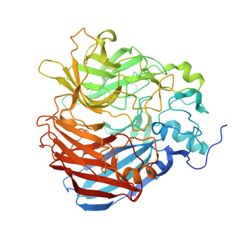
7T8P | pdb_00007t8p

CRYSTAL STRUCTURE OF T151V CAO1


Experimental Data Snapshot

  • Method: X-RAY DIFFRACTION
  • Resolution: 1.90 Å
  • R-Value Free: 
    0.204 (Depositor), 0.210 (DCC) 
  • R-Value Work: 
    0.185 (Depositor), 0.190 (DCC) 
  • R-Value Observed: 
    0.186 (Depositor) 

Starting Model: experimental
View more details

wwPDB Validation   3D Report Full Report


This is version 1.2 of the entry. See complete history


Literature

Spectroscopy and crystallography define carotenoid oxygenases as a new subclass of mononuclear non-heme Fe II enzymes.

DeWeese, D.E.Everett, M.P.Babicz Jr., J.T.Daruwalla, A.Solomon, E.I.Kiser, P.D.

(2025) J Biological Chem : 108444-108444

  • DOI: https://doi.org/10.1016/j.jbc.2025.108444
  • Primary Citation of Related Structures:  
    7T8P, 7T8Q, 8FU2, 8FU5, 8SRL

  • PubMed Abstract: 

    Carotenoid cleavage dioxygenases (CCDs) are non-heme Fe II enzymes that catalyze the oxidative cleavage of alkene bonds in carotenoids, stilbenoids, and related compounds. How these enzymes control the reaction of O 2 with their alkene substrates is unclear. Here, we apply spectroscopy in conjunction with X-ray crystallography to define the iron coordination geometry of a model CCD, CAO1, in its resting state and following substrate binding and coordination sphere substitutions. Resting CAO1 exhibits a five-coordinate (5C), square pyramidal Fe II center that undergoes steric distortion towards a trigonal bipyramidal geometry in the presence of piceatannol. Titrations with the O 2 -analog, nitric oxide (NO), show a >100-fold increase in iron-NO affinity upon substrate binding, defining a crucial role for the substrate in activating the Fe II site for O 2 reactivity. The importance of the 5C Fe II structure for reactivity was probed through mutagenesis of the second-sphere Thr151 residue of CAO1, which occludes ligand binding at the sixth coordination position. A T151G substitution resulted in the conversion of the iron center to a six-coordinate (6C) state and a 135-fold reduction in apparent catalytic efficiency towards piceatannol compared to the wild-type enzyme. Substrate complexation resulted in partial 6C to 5C conversion, indicating solvent dissociation from the iron center. Additional substitutions at this site demonstrated a general functional importance of the occluding residue within the CCD superfamily. Taken together, these data suggest an ordered mechanism of CCD catalysis occurring via substrate-promoted solvent replacement by O 2 . CCDs thus represent a new class of mononuclear non-heme Fe II enzymes.


  • Organizational Affiliation

    Department of Chemistry, Stanford University, Stanford, California 94305, USA.


Macromolecules
Find similar proteins by:  (by identity cutoff)  |  3D Structure
Entity ID: 1
MoleculeChains Sequence LengthOrganismDetailsImage
Carotenoid oxygenase 1
A, B, C, D
526Neurospora crassa OR74AMutation(s): 1 
Gene Names: cao-1NCU07008
EC: 1.13.11
UniProt
Find proteins for Q7S860 (Neurospora crassa (strain ATCC 24698 / 74-OR23-1A / CBS 708.71 / DSM 1257 / FGSC 987))
Explore Q7S860 
Go to UniProtKB:  Q7S860
Entity Groups  
Sequence Clusters30% Identity50% Identity70% Identity90% Identity95% Identity100% Identity
UniProt GroupQ7S860
Sequence Annotations
Expand
  • Reference Sequence
Small Molecules
Ligands 2 Unique
IDChains Name / Formula / InChI Key2D Diagram3D Interactions
FE2 (Subject of Investigation/LOI)
Query on FE2

Download Ideal Coordinates CCD File 
E [auth A],
G [auth B],
I [auth C],
K [auth D]
FE (II) ION
Fe
CWYNVVGOOAEACU-UHFFFAOYSA-N
CL
Query on CL

Download Ideal Coordinates CCD File 
F [auth A],
H [auth B],
J [auth C],
L [auth D]
CHLORIDE ION
Cl
VEXZGXHMUGYJMC-UHFFFAOYSA-M
Experimental Data & Validation

Experimental Data

  • Method: X-RAY DIFFRACTION
  • Resolution: 1.90 Å
  • R-Value Free:  0.204 (Depositor), 0.210 (DCC) 
  • R-Value Work:  0.185 (Depositor), 0.190 (DCC) 
  • R-Value Observed: 0.186 (Depositor) 
Space Group: P 32 2 1
Unit Cell:
Length ( Å )Angle ( ˚ )
a = 101.507α = 90
b = 101.507β = 90
c = 450.646γ = 120
Software Package:
Software NamePurpose
REFMACrefinement
PDB_EXTRACTdata extraction
XDSdata reduction
XDSdata scaling
REFMACphasing

Structure Validation

View Full Validation Report


Entry History & Funding Information

Deposition Data


Funding OrganizationLocationGrant Number
National Science Foundation (NSF, United States)United StatesCHE-2107713

Revision History  (Full details and data files)

  • Version 1.0: 2023-07-05
    Type: Initial release
  • Version 1.1: 2023-10-25
    Changes: Data collection, Refinement description
  • Version 1.2: 2025-04-09
    Changes: Database references, Structure summary