8SRL | pdb_00008srl

Crystal structure of T151V CAO1 in complex with piceatannol


Experimental Data Snapshot

  • Method: X-RAY DIFFRACTION
  • Resolution: 2.01 Å
  • R-Value Free: 
    0.191 (Depositor), 0.200 (DCC) 
  • R-Value Work: 
    0.167 (Depositor), 0.180 (DCC) 
  • R-Value Observed: 
    0.169 (Depositor) 

Starting Model: experimental
View more details

wwPDB Validation   3D Report Full Report


Ligand Structure Quality Assessment 


This is version 1.1 of the entry. See complete history


Literature

Spectroscopy and crystallography define carotenoid oxygenases as a new subclass of mononuclear non-heme Fe II enzymes.

DeWeese, D.E.Everett, M.P.Babicz Jr., J.T.Daruwalla, A.Solomon, E.I.Kiser, P.D.

(2025) J Biological Chem : 108444-108444

  • DOI: https://doi.org/10.1016/j.jbc.2025.108444
  • Primary Citation of Related Structures:  
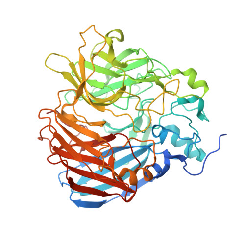
    7T8P, 7T8Q, 8FU2, 8FU5, 8SRL

  • PubMed Abstract: 

    Carotenoid cleavage dioxygenases (CCDs) are non-heme Fe II enzymes that catalyze the oxidative cleavage of alkene bonds in carotenoids, stilbenoids, and related compounds. How these enzymes control the reaction of O 2 with their alkene substrates is unclear. Here, we apply spectroscopy in conjunction with X-ray crystallography to define the iron coordination geometry of a model CCD, CAO1, in its resting state and following substrate binding and coordination sphere substitutions. Resting CAO1 exhibits a five-coordinate (5C), square pyramidal Fe II center that undergoes steric distortion towards a trigonal bipyramidal geometry in the presence of piceatannol. Titrations with the O 2 -analog, nitric oxide (NO), show a >100-fold increase in iron-NO affinity upon substrate binding, defining a crucial role for the substrate in activating the Fe II site for O 2 reactivity. The importance of the 5C Fe II structure for reactivity was probed through mutagenesis of the second-sphere Thr151 residue of CAO1, which occludes ligand binding at the sixth coordination position. A T151G substitution resulted in the conversion of the iron center to a six-coordinate (6C) state and a 135-fold reduction in apparent catalytic efficiency towards piceatannol compared to the wild-type enzyme. Substrate complexation resulted in partial 6C to 5C conversion, indicating solvent dissociation from the iron center. Additional substitutions at this site demonstrated a general functional importance of the occluding residue within the CCD superfamily. Taken together, these data suggest an ordered mechanism of CCD catalysis occurring via substrate-promoted solvent replacement by O 2 . CCDs thus represent a new class of mononuclear non-heme Fe II enzymes.


  • Organizational Affiliation

    Department of Chemistry, Stanford University, Stanford, California 94305, USA.


Macromolecules
Find similar proteins by:  (by identity cutoff)  |  3D Structure
Entity ID: 1
MoleculeChains Sequence LengthOrganismDetailsImage
Carotenoid oxygenase
A, B, C, D
526Neurospora crassaMutation(s): 1 
Gene Names: GE21DRAFT_6499
EC: 1.13.11
UniProt
Find proteins for Q7S860 (Neurospora crassa (strain ATCC 24698 / 74-OR23-1A / CBS 708.71 / DSM 1257 / FGSC 987))
Explore Q7S860 
Go to UniProtKB:  Q7S860
Entity Groups  
Sequence Clusters30% Identity50% Identity70% Identity90% Identity95% Identity100% Identity
UniProt GroupQ7S860
Sequence Annotations
Expand
  • Reference Sequence
Small Molecules
Ligands 3 Unique
IDChains Name / Formula / InChI Key2D Diagram3D Interactions
PIT (Subject of Investigation/LOI)
Query on PIT

Download Ideal Coordinates CCD File 
F [auth A],
I [auth B],
L [auth C],
O [auth D]
PICEATANNOL
C14 H12 O4
CDRPUGZCRXZLFL-OWOJBTEDSA-N
BEZ
Query on BEZ

Download Ideal Coordinates CCD File 
G [auth A],
J [auth B],
M [auth C],
P [auth D]
BENZOIC ACID
C7 H6 O2
WPYMKLBDIGXBTP-UHFFFAOYSA-N
FE2 (Subject of Investigation/LOI)
Query on FE2

Download Ideal Coordinates CCD File 
E [auth A],
H [auth B],
K [auth C],
N [auth D]
FE (II) ION
Fe
CWYNVVGOOAEACU-UHFFFAOYSA-N
Experimental Data & Validation

Experimental Data

  • Method: X-RAY DIFFRACTION
  • Resolution: 2.01 Å
  • R-Value Free:  0.191 (Depositor), 0.200 (DCC) 
  • R-Value Work:  0.167 (Depositor), 0.180 (DCC) 
  • R-Value Observed: 0.169 (Depositor) 
Space Group: P 32 2 1
Unit Cell:
Length ( Å )Angle ( ˚ )
a = 100.888α = 90
b = 100.888β = 90
c = 448.169γ = 120
Software Package:
Software NamePurpose
REFMACrefinement
XDSdata reduction
XDSdata scaling
REFMACphasing

Structure Validation

View Full Validation Report


Ligand Structure Quality Assessment 


Entry History & Funding Information

Deposition Data

  • Released Date: 2024-05-15 
  • Deposition Author(s): Kiser, P.D.

Funding OrganizationLocationGrant Number
National Science Foundation (NSF, United States)United StatesCHE-2107713

Revision History  (Full details and data files)

  • Version 1.0: 2024-05-15
    Type: Initial release
  • Version 1.1: 2025-04-09
    Changes: Database references, Structure summary