


Crystal structure of Human Prostaglandin reductase 1 (PTGR1) in complex with methotrexate (Orthorhombic C form)
Enayati, P., Lovell, S., Cooper, A., Liu, L., Battaile, K.P.To be published.
Experimental Data Snapshot
Starting Model: experimental
View more details
Entity ID: 1 | |||||
|---|---|---|---|---|---|
| Molecule | Chains | Sequence Length | Organism | Details | Image |
| Prostaglandin reductase 1 | 348 | Homo sapiens | Mutation(s): 1 Gene Names: PTGR1, LTB4DH EC: 1.3.1.48 (PDB Primary Data), 1.3.1.74 (PDB Primary Data) | 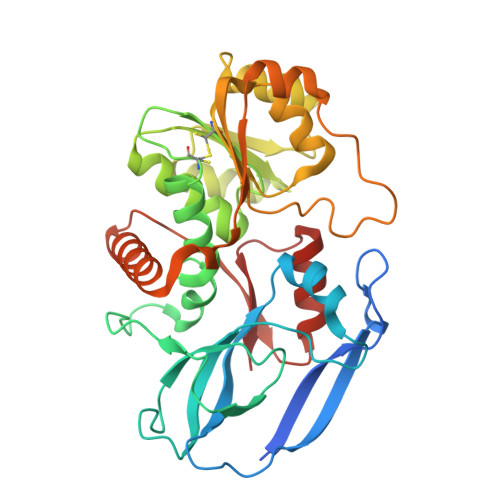 | |
UniProt & NIH Common Fund Data Resources | |||||
Find proteins for Q14914 (Homo sapiens) Explore Q14914 Go to UniProtKB: Q14914 | |||||
PHAROS: Q14914 GTEx: ENSG00000106853 | |||||
Entity Groups | |||||
| Sequence Clusters | 30% Identity50% Identity70% Identity90% Identity95% Identity100% Identity | ||||
| UniProt Group | Q14914 | ||||
Sequence AnnotationsExpand | |||||
| |||||
| Ligands 4 Unique | |||||
|---|---|---|---|---|---|
| ID | Chains | Name / Formula / InChI Key | 2D Diagram | 3D Interactions | |
| MTX (Subject of Investigation/LOI) Query on MTX | J [auth A], T [auth B] | METHOTREXATE C20 H22 N8 O5 FBOZXECLQNJBKD-ZDUSSCGKSA-N |  | ||
| PEG Query on PEG | K [auth A], U [auth B] | DI(HYDROXYETHYL)ETHER C4 H10 O3 MTHSVFCYNBDYFN-UHFFFAOYSA-N |  | ||
| DMS Query on DMS | H [auth A], I [auth A], R [auth B], S [auth B] | DIMETHYL SULFOXIDE C2 H6 O S IAZDPXIOMUYVGZ-UHFFFAOYSA-N |  | ||
| CL Query on CL | C [auth A] D [auth A] E [auth A] F [auth A] G [auth A] | CHLORIDE ION Cl VEXZGXHMUGYJMC-UHFFFAOYSA-M |  | ||
| Length ( Å ) | Angle ( ˚ ) |
|---|---|
| a = 91.473 | α = 90 |
| b = 98.209 | β = 90 |
| c = 174.444 | γ = 90 |
| Software Name | Purpose |
|---|---|
| PHENIX | refinement |
| Aimless | data scaling |
| XDS | data reduction |
| PHASER | phasing |
| Funding Organization | Location | Grant Number |
|---|---|---|
| National Institutes of Health/National Institute Of Allergy and Infectious Diseases (NIH/NIAID) | United States | 75N93022C00036 |
| National Institutes of Health/Office of the Director | United States | S10OD030394 |


