


Isoform-specific regulation of PKM by acetylation
Pavlenko, D., Tamargo-Azpilicueta, J., Ankri, Y., Nudelman, H., Shahar, A., Diez-Moreno, I., Arbely, E.(2025) Proc Natl Acad Sci U S A
Experimental Data Snapshot
Starting Model: experimental
View more details
(2025) Proc Natl Acad Sci U S A
Entity ID: 1 | |||||
|---|---|---|---|---|---|
| Molecule | Chains | Sequence Length | Organism | Details | Image |
| Pyruvate kinase PKM | 537 | Homo sapiens | Mutation(s): 0 Gene Names: PKM, OIP3, PK2, PK3, PKM2 EC: 2.7.1.40 (PDB Primary Data), 2.7.11.1 (PDB Primary Data), 2.7.10.2 (PDB Primary Data) | 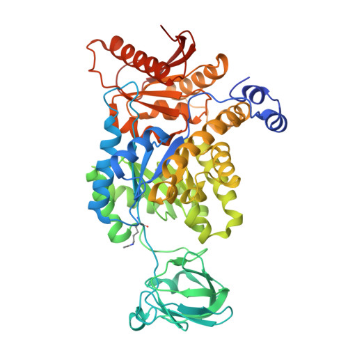 | |
UniProt & NIH Common Fund Data Resources | |||||
Find proteins for P14618 (Homo sapiens) Explore P14618 Go to UniProtKB: P14618 | |||||
PHAROS: P14618 GTEx: ENSG00000067225 | |||||
Entity Groups | |||||
| Sequence Clusters | 30% Identity50% Identity70% Identity90% Identity95% Identity100% Identity | ||||
| UniProt Group | P14618 | ||||
Sequence AnnotationsExpand | |||||
| |||||
| Ligands 5 Unique | |||||
|---|---|---|---|---|---|
| ID | Chains | Name / Formula / InChI Key | 2D Diagram | 3D Interactions | |
| FBP (Subject of Investigation/LOI) Query on FBP | E [auth A], K [auth B], O [auth C], U [auth D] | 1,6-di-O-phosphono-beta-D-fructofuranose C6 H14 O12 P2 RNBGYGVWRKECFJ-ARQDHWQXSA-N |  | ||
| SIN (Subject of Investigation/LOI) Query on SIN | J [auth A], N [auth B], Q [auth C], W [auth D] | SUCCINIC ACID C4 H6 O4 KDYFGRWQOYBRFD-UHFFFAOYSA-N |  | ||
| GOL (Subject of Investigation/LOI) Query on GOL | P [auth C], R [auth C] | GLYCEROL C3 H8 O3 PEDCQBHIVMGVHV-UHFFFAOYSA-N |  | ||
| EDO (Subject of Investigation/LOI) Query on EDO | F [auth A] G [auth A] H [auth A] I [auth A] L [auth B] | 1,2-ETHANEDIOL C2 H6 O2 LYCAIKOWRPUZTN-UHFFFAOYSA-N |  | ||
| K (Subject of Investigation/LOI) Query on K | S [auth C], T [auth C] | POTASSIUM ION K NPYPAHLBTDXSSS-UHFFFAOYSA-N |  | ||
| Modified Residues 1 Unique | |||||
|---|---|---|---|---|---|
| ID | Chains | Type | Formula | 2D Diagram | Parent |
| ALY Query on ALY | A, B, C, D | L-PEPTIDE LINKING | C8 H16 N2 O3 |  | LYS |
| Length ( Å ) | Angle ( ˚ ) |
|---|---|
| a = 81.229 | α = 90 |
| b = 155.169 | β = 107.61 |
| c = 101.023 | γ = 90 |
| Software Name | Purpose |
|---|---|
| REFMAC | refinement |
| XDS | data reduction |
| Aimless | data scaling |
| PHASER | phasing |
| Funding Organization | Location | Grant Number |
|---|---|---|
| European Research Council (ERC) | European Union | 678461 |
| Israel Science Foundation | Israel | 1769/20 |


