


Structure of ALAS bound to succinyl-CoA from S. cerevisiae
Tran, J.U., Brown, B.L.To be published.
Experimental Data Snapshot
Starting Model: experimental
View more details
Entity ID: 1 | |||||
|---|---|---|---|---|---|
| Molecule | Chains | Sequence Length | Organism | Details | Image |
| 5-aminolevulinate synthase, mitochondrial | A [auth C], B [auth D], C [auth A], D [auth E], E [auth B] | 491 | Saccharomyces cerevisiae | Mutation(s): 0 Gene Names: HEM1, CYD1, YDR232W, YD9934.16 EC: 2.3.1.37 |  |
UniProt | |||||
Find proteins for P09950 (Saccharomyces cerevisiae (strain ATCC 204508 / S288c)) Explore P09950 Go to UniProtKB: P09950 | |||||
Entity Groups | |||||
| Sequence Clusters | 30% Identity50% Identity70% Identity90% Identity95% Identity100% Identity | ||||
| UniProt Group | P09950 | ||||
Sequence AnnotationsExpand | |||||
| |||||
Entity ID: 2 | |||||
|---|---|---|---|---|---|
| Molecule | Chains | Sequence Length | Organism | Details | Image |
| 5-aminolevulinate synthase, mitochondrial | 491 | Saccharomyces cerevisiae | Mutation(s): 0 Gene Names: HEM1, CYD1, YDR232W, YD9934.16 EC: 2.3.1.37 | 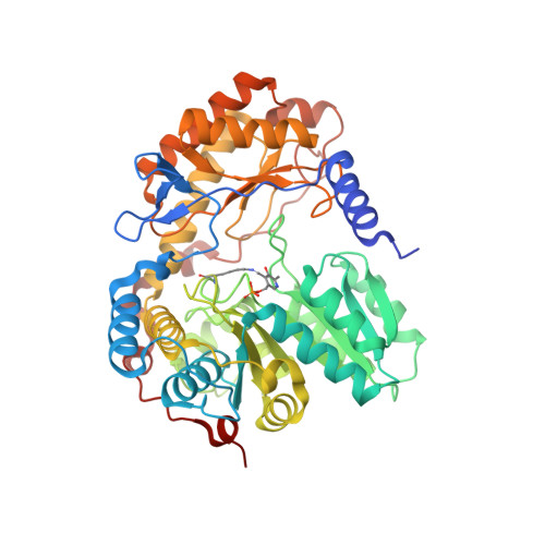 | |
UniProt | |||||
Find proteins for P09950 (Saccharomyces cerevisiae (strain ATCC 204508 / S288c)) Explore P09950 Go to UniProtKB: P09950 | |||||
Entity Groups | |||||
| Sequence Clusters | 30% Identity50% Identity70% Identity90% Identity95% Identity100% Identity | ||||
| UniProt Group | P09950 | ||||
Sequence AnnotationsExpand | |||||
| |||||
| Ligands 3 Unique | |||||
|---|---|---|---|---|---|
| ID | Chains | Name / Formula / InChI Key | 2D Diagram | 3D Interactions | |
| SCA (Subject of Investigation/LOI) Query on SCA | H [auth C], L [auth A], N [auth E], P [auth B], Q [auth F] | SUCCINYL-COENZYME A C25 H40 N7 O19 P3 S VNOYUJKHFWYWIR-ITIYDSSPSA-N |  | ||
| PLP (Subject of Investigation/LOI) Query on PLP | G [auth C], I [auth D], K [auth A], M [auth E], O [auth B] | PYRIDOXAL-5'-PHOSPHATE C8 H10 N O6 P NGVDGCNFYWLIFO-UHFFFAOYSA-N |  | ||
| MG Query on MG | J [auth D] | MAGNESIUM ION Mg JLVVSXFLKOJNIY-UHFFFAOYSA-N |  | ||
| Modified Residues 1 Unique | |||||
|---|---|---|---|---|---|
| ID | Chains | Type | Formula | 2D Diagram | Parent |
| LLP Query on LLP | F | L-PEPTIDE LINKING | C14 H22 N3 O7 P |  | LYS |
| Length ( Å ) | Angle ( ˚ ) |
|---|---|
| a = 60.595 | α = 116.403 |
| b = 114.241 | β = 97.507 |
| c = 119.307 | γ = 91.839 |
| Software Name | Purpose |
|---|---|
| REFMAC | refinement |
| XDS | data reduction |
| XDS | data scaling |
| PHASER | phasing |
| Funding Organization | Location | Grant Number |
|---|---|---|
| National Institutes of Health/National Institute of General Medical Sciences (NIH/NIGMS) | United States | DP2 GM146255 |
| American Heart Association | United States | 24PRE1190012 |


